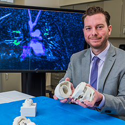
Through my interest in congenital cardiac MRI, I have teamed up with bioengineers at the Jump Trading Simulation & Education Center to create exact-replica models of children’s hearts. We’ve been on this journey in which we are creating processes that have never existed before, and the innovative part of the Jump lab is this mindset that if it doesn’t exist, we will create it.
It’s been very fun to be part of this project in which we produce a 3D heart model from 2D images. Over the past year, we’ve been refining our process. We’ve had challenges, but successes as well. We’ve now refined it down to where we can fairly quickly take a heart out of an MRI and literally put a model of it into our hands.
Benefits for Surgery
With 3D printing, surgeons can make better decisions before they go into the operating room. The more prepared they are, the better decisions they make, and the fewer surprises they encounter. Surgeons typically rely on 2D images taken by ultrasound, MRI and special x-rays called CT scans to plan their surgeries, but these images may not reveal the complex structural defects present in a patient. Using the 2D images to create 3D models, even out of simple, plaster-like materials, has provided very valuable information that wasn’t available before. When you’re holding the heart model in your hands, it provides a new dimension of understanding that you just don’t get from looking at a screen.
For example, I recently had two requests for patients that already had MRIs for the heart models, and by the end of the week, we had the models printed. It’s a tool our surgeons are beginning to rely on. Not long ago, I was down in the operating room taking a model to the surgeon because he had a question prior to surgery.
The beauty of the project is that children with complex heart disease may have only half of a heart to work with, so when you’re trying to figure out the optimal operation, there’s no better way than to actually see the heart and hold it in your hands before attempting the surgery. You can see that if you make a compromise here, that would improve, or fix this other problem. The approach is still new—3D printing has not been approved by the U.S. Food and Drug Administration—and our study is small, but the findings thus far have been nothing but positive.
The NIH 3D Print Exchange
While we are using these models for actual surgical planning—which I believe is a tremendous, incredible use for our patients—there are also a lot of opportunities to share this information with the world in a type of library format.
One issue we’ve identified is that there aren’t anatomic models that describe congenital heart disease or all the variations; it’s all been limited to pictures and textbooks. To be able to actually hold that model in your hand is a new kind of library that would allow more people to learn from what we are doing and have done—that is what we’re striving to achieve.
The logistics of creating the architecture and the ability to support the library online were going to be big hurdles. Luckily, the National Institutes of Health 3D Print Exchange happened to come to life at the exact right time. We have been very impressed with its infrastructure, but more importantly, its mindset. In speaking with Dr. Darrell Hurt at the NIH, it’s clear he understands the significance of taking something you’ve understood for years, printing it out, and holding it in your hands. This method can provide a whole new level of understanding. That’s exactly how we feel about these hearts.
For example, take something as simple as an artrial septal defect (ASD)—something I’ve read about in textbook after textbook and seen in thousands of echocardiograms. When we printed out an ASD and I was able to hold it in my hand for the first time, I learned something new. We’ve begun the process of taking these perfect examples of pathology, where we have great images, and creating 3D models that we’re now able to put on the NIH 3D Print Exchange. This will allow anybody to go online, download the file, and if you have a 3D printer, print it out. This includes medical schools, hospitals, physicians, high schools and beyond.
Establishing a Collaborative Effort
We hold ourselves to a very high quality-assurance standard because we want people to learn and use these models as training tools. Our next step is to elevate the project by working with a collaborative group of people to create a peer-review system that provides very accurate anatomic examples of congenital heart disease. We hope to take the benefits of the 3D modeling resources available here at Jump and make them available to anybody willing to learn through the work the NIH is doing with the 3D Print Exchange.
Right now, we have several hearts up on the website—two pathologic specimens that were designed for the library. They come from patients with ideal images that demonstrated the specific anatomic detail. We have also included a brief description file on the website. Also on the Print Exchange are some of our research hearts, which were designed to show certain elements that are useful in surgeries but are not fully detailed.
By having these hearts on the 3D Print Exchange as we move forward, we will be able to reference them when we present at scientific meetings. They also provide a reference point if we decide to conduct research with those specific hearts. I believe the work the Print Exchange is doing is going to explode overnight at some point. There’s a lot of potential there, and while the project is still in its infancy, it contains tremendous molecular biology. We really see a lot of potential for the future of the NIH 3D Print Exchange and medical or anatomic libraries. iBi
Dr. Matthew Bramlet is a pediatric cardiologist and assistant professor of pediatrics at the University of Illinois College of Medicine at Peoria.

