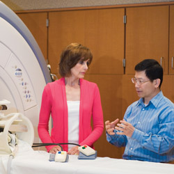
Still in its infancy, the Center for Collaborative Brain Research (CCBR), a joint project of Bradley University, Illinois Neurological Institute (INI), OSF Saint Francis Medical Center and the University of Illinois College of Medicine at Peoria (UICOMP), encourages a new, community-wide collaboration.
“We are a very lucky community in that we have a very strong neurology and neurosurgery presence in this area,” explains Tom Cox, CCBR board member and director of radiology at OSF Saint Francis Medical Center. “Not all cities this size have that.”
Founded in March 2010 by co-directors Dr. Lori Russell-Chapin of Bradley University and Dr. Wen-Ching Liu of OSF, the Center strives to create an ongoing network between the groups to elevate support of clinical diagnosis and treatment by improving current academic inquiries in brain research, neural feedback and brain imaging.
“We all stick to our own silos and specific disciplines, and I just always think, ‘Wow, I wonder if it would be better if we all worked together conjointly,’” says Russell-Chapin. “It makes a lot more sense—we each bring our own area of expertise and it makes [the work] richer.”
“Let’s Do It!”
“Each group has its own strength,” Liu agrees. As a clinical physicist and the director of functional magnetic resonance imaging (fMRI) at OSF Saint Francis Medical Center, his department provides imaging for the CCBR’s various research projects. Meanwhile, the INI contributes to projects from a clinical standpoint; Bradley and UICOMP offer academic expertise. “We’re trying to provide an integration of all these different people…to provide a more complete answer,” he says.
The CCBR began with a conversation between Dr. Liu and Tom Cox. “We were talking about how we can better promote our department and the new cutting edge fMRI ability we have,” Liu explains.
“We felt that our fMRI program was an underutilized tool, so we wanted to expose more people to what it can do,” adds Cox.
What functional MRI can do is offer visual evidence of functioning inside the brain—the “best way to look into the brain without opening [it] up,” Liu notes. Using a magnetic field, radio frequency pulses and a computer, fMRI can produce detailed images of nearly any internal body structure, but its unique access to the brain tops the list. Images of the brain can be collected and analyzed to examine its anatomy; determine which parts handle which functions; assess effects of stroke, trauma or disease on performance; monitor tumor growth; plan for brain surgery; and more.
“A lot about the brain hasn’t been answered, and it’s hard to answer everything doing behavioral studies,” stresses Liu. “The information we get out of some of these studies will lead to—we’re hoping—fMRI becoming more of an integral part of the diagnosis and treatment of disease than it is now.”
Eager to share fMRI’s prospects with area researchers, Dr. Liu visited Bradley’s campus and delivered a lecture on brain imaging’s scope beyond clinical use. Dr. Russell-Chapin was in the room. “Let’s do this!” Liu recalls her saying, enthusiastic about the idea of combining forces from the get-go.
Pilot Projects Demonstrate Teamwork
“Truly, the center allows Bradley to do research that we would never be able to do otherwise,” explains Russell-Chapin. “I was really pleased with Peoria for knowing that collaboration is the way to go. Bradley shouldn’t sit by itself, OSF shouldn’t sit by itself. I shouldn’t sit by myself… In fact, I had eight students helping me in my [pilot project] research from all different disciplines: nursing, psychology, biology and counseling. Those students never get together otherwise. How much fun is that?”
Her recently completed project—and the Center’s first pilot study—explored the ways in which real-time neurofeedback can be used to re-regulate the brain in children diagnosed with ADHD. In the fMRI scans of the pre- and post-tests of patients in the treatment group, both the precuneus, a coil in the parietal lobe of the brain involved in memory, coordination and visual-spatial ability, and the default mode network, the brain region active when an individual is not focused, appear consolidated after treatment. The CCBR, she explains, allows researchers to take their own area of expertise, fantasize about how the ability to look into the brain might help them in that research, then actually do it—whether it’s examining its anatomy more closely, or in her case, receiving visual evidence of functioning improvement through proposed treatment.
 The CCBR accepts proposals, like Dr. Russell-Chapin’s, which demonstrate a desire to collaborate at each of the Center’s levels. “We typically will not take a research project that does not show collaboration. It’s called the Center for Collaborative Brain Research, so we expect that there will be a Bradley or UICOMP component, medical staff involvement and an imaging component,” says Cox.
In another example, a pilot project is underway by psychologist Dr. Allen I. Huffcutt at Bradley University to explore the patterns of brain activity in response to employment interview questions. With the paradigm built and dummy testing now complete, the subject interviews will begin soon “to see what part of the brain lights up when we’re asking targeted questions,” explains Russell-Chapin, co-investigator of the project.
Everyone Benefits
“The CCBR benefits Bradley greatly because we get to expand the diversity of research with the fMRI…, offer more courses in neuroscience…[and] talk more amongst varying disciplines.” Russell-Chapin says. “But I think it’s also a community-based benefit.”
“We are creating relationships that I really do think the community will benefit directly from,” says Cox. “Not every one of our projects uses functional MRI, but I’d say the lion’s share do. [The CCBR] has enhanced awareness within the medical community of what the tool can do… The more we do in that regard, the more it will be integrated for use in the community.” iBi

