Advanced Technology to Diagnose Lung Disease

An endobronchial ultrasound (EBUS) procedure is an efficient, accurate way to investigate suspicious lymph nodes in the lung. The advanced technology used in this procedure provides the trained pulmonologist an internal perspective of the afflicted area and allows for an on-the-spot biopsy if necessary.
“The advanced technology of an EBUS procedure has clear advantages over mediastinoscopy, which is generally used to sample thoracic lymph nodes: it’s less invasive, does not require general anesthesia and still provides an accurate diagnosis,” says Dr. Patrick Whitten, an interventional pulmonologist with the Illinois Lung Institute and member of the OSF Saint Francis team committed to creating a Lung Center of Excellence.
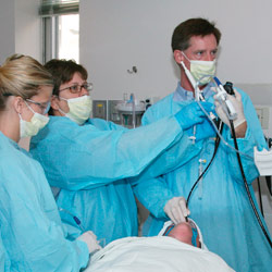
He also credits procedure efficiency to the strength of his team, which includes: a sedation nurse; an airway and hemodynamic monitoring nurse who monitors the oxygen level, heart rate and blood pressure to maintain appropriate levels; an ultrasound/radiology technician; and a cytology team for onsite tissue processing and evaluation. “The depth of knowledge and the concentrated practical application of the team ensures that the procedure runs smoothly.”
OSF Saint Francis has been utilizing EBUS technology for four years and is currently the only medical center in central Illinois that is performing procedures with this equipment. The majority of medical centers do not have a focused team for the treatment of lung disease, making the equipment a lesser priority and cost-prohibitive.
The EBUS Procedure
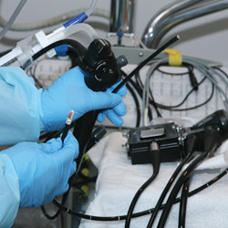
At the onset of the procedure, the patient is put into a conscious sedation. A pulmonologist inserts a scope down the throat and guides it to the appropriate area through direct inspection. Once it has reached the targeted area, ultrasonography provides an illuminated visualization in actual time. Both lymph node tissue and vessels can be identified. A needle aspiration of the node or lesion is performed for tissue acquisition and sampling if the afflicted area warrants further investigation. Tiny forceps may be used if necessary to provide a larger tissue sample. All in all, this advanced procedure takes longer to prep than to complete! How Does it Differ From the Typical Procedures?
Typical bronchoscopy can only see in the airway lumen itself. If lymph nodes are to be sampled, it is done blindly since they arise outside the airway lumen. Ultrasound bronchoscopy allows for direct visualization of the lymph node for accurate sampling, thus creating a safer way to obtain tissue.
An EBUS procedure takes approximately the same time as a traditional bronchoscopy (generally 30 minutes) and is far less invasive than other procedures performed to sample lymph nodes in the chest.
As opposed to mediastinoscopy, EBUS does not require general anesthesia and the patient experiences less recovery pain because no incisions are made. The patient is still able to get an accurate diagnosis, which empowers him or her to move out of the stressful state-of-not-knowing and into a more action-oriented position. If the diagnosis is cancer, the more responsive diagnostic time allows the patient to seek treatment options quicker.
Why Create a Lung Center of Excellence?
Lung disease has been on the rise, increasing 220 percent from the 1970s through the 1990s. Lung cancer is the number one cancer killer in women, and approximately 12 percent of all lung cancer occurs in non-smokers.
Dr. William Tillis was responsible for bringing EBUS technology, as well as other cutting-edge bronchoscopic technologies, such as Electromagnetic Navigation Bronchoscopy—a GPS-like technology that allows more precise tracking and treatment of small lesions on and around the lung—to OSF Saint Francis when he recognized the increasing need for treatment of lung disease in central Illinois. After receiving administrative approval, he worked with the OSF Foundation to purchase this equipment and train physicians.
Creating a Lung Center of Excellence means assembling a team of specialists whose knowledge and expertise focus specifically on the human lung and equipping them with the most advanced tools. Not only does this provide patients with a higher standard of care associated with specialization, it also creates a best-in-class resource for training physicians to deliver similar care throughout the country. iBi

