
Mammography Goes Digital
For women, breast cancer is the most common non-skin cancer and the second leading cause of cancer-related deaths in the U.S. The ongoing battle against breast cancer moved ahead this month with central Illinois’ first all-digital mammography imaging department at Methodist Medical Center.
Just as consumers today have to decide between analog and digital television sets, so, too, do physicians and radiology centers have to choose between analog and digital mammography units.
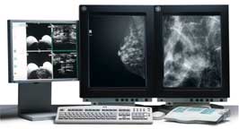
Like analog mammography, digital mammography is basically an X-ray of the breast used for both screening and diagnosis. But unlike traditional mammography, which uses film as both a receptor and a display for the image to produce fixed images, digital mammography uses detectors—similar to those found in digital cameras—that change the X-rays into electrical signals. These signals then are transferred to a digital receptor that converts the X-ray energy to numbers, processes the numbers, and produces an image that can be displayed on a monitor or printed on a high-resolution laser printer. Because there’s a lower level of intrinsic noise, a lower dose of X-ray may be used than currently is used in conventional mammography.
Digital mammography also helps provide more accurate breast cancer diagnoses that may, in turn, reduce unnecessary invasive biopsies. That makes the decision to create a digital mammography center a logical step in improving health care delivery for women.
Studies have found several other advantages to digital mammography systems, including:
• Improved detection because digital mammography can distinguish more shades of gray than film mammography.
• Digital mammography provides the capability to acquire images in near-real time and to process them quickly and accurately.
• Digital imaging provides the ability to archive films electronically. This not only reduces the risk of misplacing or damaging films, but provides for immediate electronic transmittal via the Internet for a second opinion, regardless of where the physician is located.
• Digital exams take less than half the time of conventional, film-based mammograms, with a digitally displayed image available in about 10 seconds, compared to two minutes for a conventional film mammogram, which then must be developed.
• Digital images result in fewer patient callbacks because of the ability to generate images within seconds and the flexibility to magnify and manipulate those images.
The GE Senographe 2000 full field digital mammography system is designed to meet a broad spectrum of clinical needs, from screening and diagnosis to interventional procedures. The new GE systems are designed to image the breasts in both female and male patients. The systems also utilize the R2 computer-aided program that provides radiologists with a “second pair of eyes” when reading a mammogram. Similar to a spellchecker system on a personal computer, this technology has the potential to detect findings that otherwise might be overlooked during the review process, thus improving the opportunity for cancer detection. TPW